Diagnosis
Triple assessment is the key element of diagnosis of breast cancer. It comprises the following clinical examination,breast imaging and a tissue diagnosis with a biopsy. Combined results of the three tests does increase the accuracy of the diagnosis.
Clinical Examination
The doctor will take a thorough history to assess your risk of breast cancer and then examine your breasts and axilla (armpit).
Imaging
Mammogram requires compression of the breast between two plates and is uncomfortable. Two views are usually obtained . With modern equipment the radiation dose is negligible. The benefit from having your mammogram far outweighs its risks. Women with implants can safely have a mammogram. For more information: www.cancerscreening.gov.au

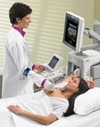
Ultrasound may also be used in diagnosing breast lesions. It is painless and well tolerated by the patient. It is particularly helpful in younger women under the age of 40.
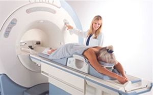
After analyzing your family history and current problem your breast surgeon may recommend a medicare non-rebatable MRI. Of course this will be discussed with you.

Biopsy
Depending on outcome of your clinical examination and imaging results you may be advised to have :
- Fine Needle Aspiration Cytology
- Under image guidance, a fine needle is introduced into the lesion and small amount of cells are withdrawn for examination. This procedure usually does not require anaesthetic. This is a study of cells only and will require an expert cytologist to analysis the results.
- Core Biopsy
- Open Surgical Biopsy
The disadvantage is THAT it cannot differentiate in-situ from invasive cancers and may yield a non-diagnostic result.
Under image guidance, local anaesthetic is used to introduce a much larger needle to remove tissue for examination. A larger sample is obtained compared to a fine needle cytology and more information is obtained .
The breast surgeons would like to obtain information prior to planning surgery. However in a small group of patients, the FNAC and Core biopsy is not able to obtain a definite diagnosis. These patients will require an open surgical biopsy.
This is performed under general anaesthetic usually as a day care procedure. Minimal (10-20gm) of breast tissue is removed. Occasionally a small wire may be introduced to guide the surgeon.
Staging
The investigations may include a CT scan of the brain, chest, abdomen and pelvis. A bone scan may also be undertaken.

 Gosford


